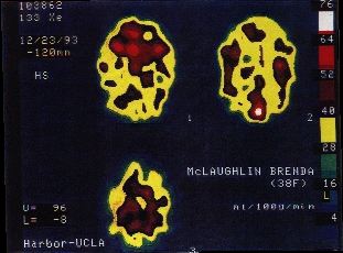
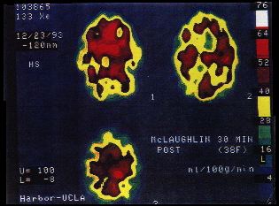
Single Photon Emission Computerized Tomography (SPECT) Scan of MCS Patient's Brain Before and After Challenge with Perfume Inhalation
(NOTE: Text in the following report is reproduced for clarity.)
DOCTORS REPORT:
HARBOR UCLA DIAGNOSTIC IMAGING CENTER
HARBOR UCLA PROFESSIONAL OFFICE BUILDING
21840 SOUTH NORMANDIE AVENUE
TORRANCE, CALIFORNIA 90502
APPT. (213)212-5939 – NUCLEAR MEDICINE DEPT.
213-783-5273
BRAIN SPECT REPORT
PATIENT NAME: McLaughlin, Brenda
EXAM DATE: December 23, 1993
AGE: 38
SEX: F
NO: 2688
REFERRING PHYSICIAN: (removed at physician's request)
Exam
This concerns your patient of the above name referred for evaluation of cerebral perfusion. On 12/23/93 regional cerebral blood flow by means of Xe-133 was determined with the Shimadzu Brain Dedicated Unit. After inhalation of 30mCi of Xe-133, cerebral blood flow was found to fluctuate between 40 ml/min/100g in both dorsal frontal lobes and both temporal lobes with focal hypoperfusion also in both posterior parietal lobes. Maximum perfusion is observed in the visual cortex at 76 ml/min/100g and the remainder of the gray matter blood flow fluctuates between 40 and 64 ml/min/100g.
Thirty minutes post inhalation of perfume with PetCO2 of 32 vs. 35 ml/min/100g, there is an increase perfusion in the dorsal aspects of the frontal lobes from 40 to 60 ml/min/100g with an increase of perfusion also in the right temporal lobe. Maximum perfusion is 64 ml/min/100g in the basal ganglia and the remainder of the gray matter blood flow fluctuates between 40 and 64 ml/min/100g.
The study was followed by brain SPECT by means of HMPAO (Ceretec, Amersham) for assessment of regional cerebral perfusion. 60 min. after IV injection of 30 mCi of HMPAO, nine 1.6 cm. thick transaxial slices of the brain are acquired and a 3-dimensional image is reconstructed by back projection with adequate filtering. Coronal and sagittal frames are displayed, demonstrating bilateral temporal hypoperfusion, bilateral dorsal frontal hypoperfusion with diminution of perfusion in the dorsal cingulate gyrus and marked thinning of cortical perfusion in dorsal aspects of frontal and parietal lobes with a pattern of scalloping.
In conclusion, findings suggest: 1) Diminished cerebral blood flow. 2) Bilateral frontal, temporal and parietal hypoperfusion. 3) Marked scalloping pattern of perfusion in frontal and parietal lobes. 4) Vasculitis vs. exposure to neurotoxic substances.
I thank you for referring Ms. McLaughlin to our laboratory.
Sincerely yours,
(Personal info removed at request of physician.)


